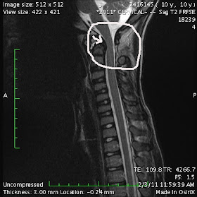
Here are pictures from Axel's MRI done a couple weeks ago. I put an arrow to the problem areas. It is important to remember when looking at these that Axel did not have one single symptom!!!! As his surgeon put it, he was "one bad cough away from catastrophic injury".
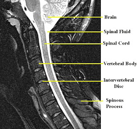
First, here is a picture of a normal Cervical Spine MRI. You can see how the spinal cord maintains it's shape up the entire spinal column, and there is a pretty equal amount of spinal fluid around it all the way up the length as well.
This first picture shows a couple of things. First, the arrow points to where the C1 vertebra is pushing against the spinal cord. Also, you can see on the front (left) side of the cord, between the C2 and C3 vertebrae, the disk looks different that the disks between the other vertebrae. That's because it doesn't have enough fluid in it to provide a cushion between the two vertebrae.
This pictures shows the Cerebrospinal Fluid (CSF) that surrounds the spinal cord. It is the white area around the cord. You can see where the arrow is how the CSF fluid is being blocked from getting to the brain. Thankfully it can also get to the brain on the back side of the spinal cord as well. Also, the spinal cord itself is more narrow in that area because of the movement of the C1 vertebrae.


So what are they going to do to help him? You all will be in my prayers.
ReplyDeleteHe'll be having a spinal fusion done the end of March. This means a surgery 4-6 hour surgery, then a halo for the next 6 months after surgery.
ReplyDeleteNow in see. Thanks for the explanation.
ReplyDeleteWow! You guys will be in our prayers the next few weeks.
ReplyDeleteYes, thinking of you ... its always scary going into hospital, but most kids seem to take it in their stride. Its the grown ups who get anxious - ignorance is bliss, eh?
ReplyDeleteWow, this post showed up on a google search for "white spots on cervical MRI". I was like...hey! I know them! And now he's all good, right? :D Perhaps I will be so blessed...
ReplyDeleteWas he diagnosed with cranio-cervical instability or Basilar Invagination? I HIGHLY RECOMMEND getting a brain to pelvis MRI with and without contrast to check for Syringomyelia. ALSO Ehlers-Danlos Syndrome(EDS) needs to be looked into! EDS is a connective tissue disorder that was first recognized by Hippocrates yet medical professionals are generally unfamiliar with it. EDS III-HYPERMOBILITY Type is based upon family pedigree and clinical diagnosis based on the Beighton-scale. The other forms are generally diagnosed through genetic testing. EDS IV-Vascular Type can be fatal. ALSO people with EDS often do not respond to antidepressants, benzos, opiates, local anesthesia, and other medications as they 'should.' More often than not we require a much higher dosages of medications. EDS is a connective tissue disorder that causes moderate to severe, often debilitating, pain due to reoccurring sublaxations and dislocations. If there is no diagnosis it is assumed we are pill seekers trying to get a high. Steroids should NEVER EVER be administered unless it is a life threatening situation bc steroids break down collagen which causes further joint instability.
ReplyDeleteI hope I was some sort of help.
www.wstfcure.org is a great site
Thank you for your comment Brook. AAI occurs in approximately 10% of those who have Down syndreom (Axel has DS) He does not have EDS, and he responds well to both local and general anesthetics. Also, this bost is now 6 years old. Axel is long since recovered from his spinal fusion with no long lasting effects or damage.
ReplyDeleteThank you for your comment Brook. AAI occurs in approximately 10% of those who have Down syndreom (Axel has DS) He does not have EDS, and he responds well to both local and general anesthetics. Also, this bost is now 6 years old. Axel is long since recovered from his spinal fusion with no long lasting effects or damage.
ReplyDelete